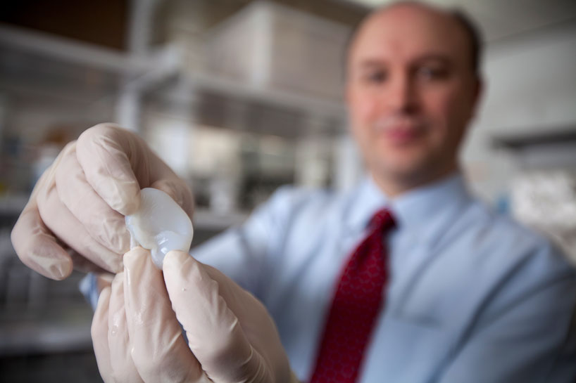
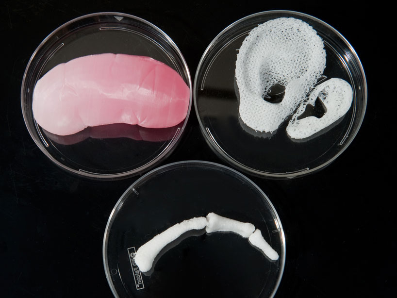
scientists at cornell university 3D print artificial earsimage © lindsay france


in the most recent medical development in artificial organ creation, a team of scientists and physicians from cornell university have engineered 3D printed life-like ears used to treat children born with a congenital deformity called microtia, or help individuals who have lost part or all of their external ear in an accident or from cancer. the artificial body tissue is made by processing additive gels found within living cells – with collagen derived from rat tails and cartilage taken from cow ears.


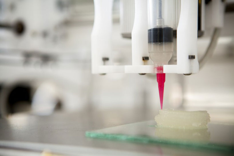
the modelling process is digitized into an assemble-able human ear, where then high-density gel, similar to the consistency of jell-o is injected into a mold – resulting in flesh resembling that of a human-being.in the future, the engineers will look at new ways to develop populations of human ear cartilage cells from patients instead of cow cartilage, which would reduce any possibility of rejection.


lawrence bonassar, associate professor of biomedical engineering adds: ‘it takes half a day to design the mold, a day or so to print it, 30 minutes to inject the gel, and we can remove the ear 15 minutes later. we trim the ear and then let it culture for several days in nourishing cell culture media before it is implanted.’


3D printed organs from regenerative living cells

injection processimage © lindsay france


early stages of bio-printing ears at cornell universityimage courtesy BBC


3D printed organs from regenerative living cells

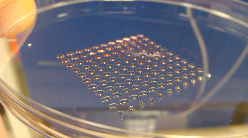
3D printed embryonic stem cellsimage courtesy heriot-watt university


a 3D printing technique developed by scientists at the heriot-watt university in edinburgh produces clusters of human embryonic stem cells – a breakthrough which could pave the way to purpose-built replacement organs for patients, eliminating the need for organ donation, immune suppression and the problem of transplant rejection. the technique will enable more accurate human tissue models to be created – which are essential for drug development and toxicity-testing.


3D printed organs from regenerative living cells

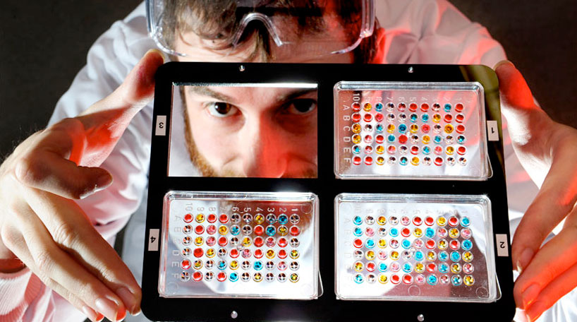
artificial samples of ‘livers on a chip’ image courtesy heriot-watt university


the team, led by heriot-watt university’s dr. will shu is also working towards creating the world’s first artificial human livers for drug testing, drastically cutting the amount of live animals used and getting life-saving medicines into the clinic faster. the process aims to develop miniature human liver tissues – where eventually ‘livers-on-a-chip’, could be implemented using technology that will ‘print’ the cells in three-dimension onto testing surfaces.


3D printed organs from regenerative living cells

regenerative livers grown with 3D printersimage courtesy wake forest institute


in the earliest technological investigation, physicians and scientists at the wake forest institute for regeneration were the first in the world to conduct and engineer laboratory-grown organs that were successfully implanted into humans. currently, the team is working towards engineering more than 30 different replacement tissues and organs – includingbladder, trachea, cartilage and heart healing therapies to cure disease. since 2006, cases such as implanted bladders, urine tubes and spinal cords have been successfully transplanted into patients, with long term success being reported in the clinical trials.


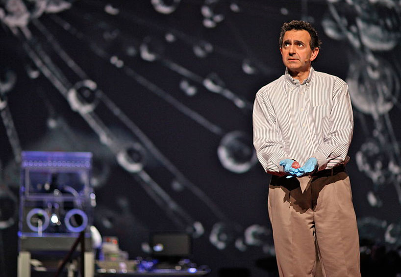
 3D printed organs from regenerative living cellsanthony atala, professor and director of wake forest institute for regenerative medicine – holding a 3D printed kidney during his TED talk in 2011



anthony atala ‘s TED talkvideo courtesy TED


engineering tiny livers in the labvideo courtesy WFIFR